Which lesions need assessment?
Appearance alone cannot always establish whether a skin growth is benign or malignant. Growing, bleeding, nonhealing or changing lesions need assessment. Basal cell carcinoma, squamous cell carcinoma and melanoma require different diagnostic and treatment plans. Some benign lesions are removed for irritation or uncertainty. Excision aims to obtain an appropriate specimen and treatment margin, not merely erase a visible bump.
Examination and biopsy planning
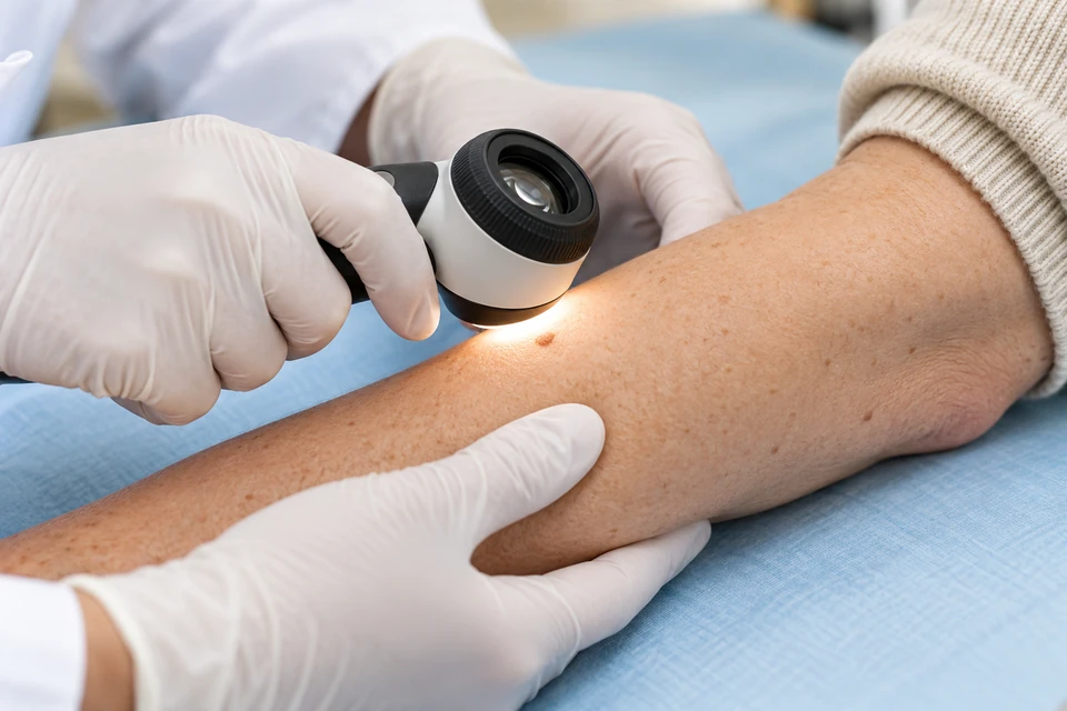
Assessment reviews duration, change, bleeding, sun exposure, previous skin cancer and immune suppression. Dermatological examination and dermoscopy may help. Biopsy type depends on location and suspicion; possible melanoma requires sampling that permits meaningful depth assessment. Clear site documentation assists later treatment. Lesions near sensitive facial structures or functionally important areas may need additional specialist input.
Alternatives to standard excision
Selected confirmed superficial, low-risk lesions may suit topical treatment, freezing or other methods. These are not interchangeable options for every suspicious growth. Destroying a possible melanoma without a suitable specimen can impair diagnosis. Mohs or staged margin-controlled surgery may suit certain high-risk or sensitive-site cancers. Radiotherapy is another selected option. Diagnosis, recurrence risk, function and operative suitability guide choice.
Excision and wound closure
Excision is often performed under local anaesthesia, with alternatives for larger or difficult procedures. Tissue is removed with diagnosis-specific margins and sent for pathology. Closure may use stitches, open healing, a flap or a skin graft. Reconstruction can be coordinated with margin assessment. Removing the visible lesion does not guarantee clear microscopic margins; further treatment may still be required.
Risks and the importance of pathology
Risks include bleeding, infection, wound separation, scarring, colour change, numbness and injury to nearby structures. Flaps or grafts add risks of incomplete survival and extra care. Pathology establishes tumour type and relevant depth, margins and risk features. Melanoma may require further excision or selected lymph-node assessment, but these are not necessary for every skin lesion.
Preparation and follow-up
Report anticoagulants, allergies, implanted devices and healing conditions, without independently stopping medicines. Follow dressing, washing, stitch and tension-protection instructions. Activity depends on wound location and closure. Confirm how pathology will be communicated. A cancer diagnosis leads to risk-based follow-up and skin checks. Sun protection and recognising new changes remain part of care after the wound has healed.
Early and later warning signs
Seek help for persistent bleeding despite instructed measures, rapidly increasing swelling, spreading redness, fever or pus. Marked colour change in a graft or flap or increasing pain should be reported quickly. Breathing difficulty or fainting requires emergency assessment. Later, a new nodule or nonhealing area at the scar needs review. Do not assume an uncommunicated pathology result is normal.
This information is general. The cause of symptoms, appropriate investigations and treatment options are determined through individual assessment.