Why a lymph node may need examination
Lymph nodes are part of the immune system and can enlarge for benign reasons, including infection. Enlargement does not automatically mean cancer. Persistent, growing or unexplained nodes may need tissue sampling after assessment. Excisional biopsy generally removes a selected whole node to examine its architecture. This is usually diagnostic and differs from a therapeutic lymph-node dissection involving a larger group.
Assessment and selecting the node
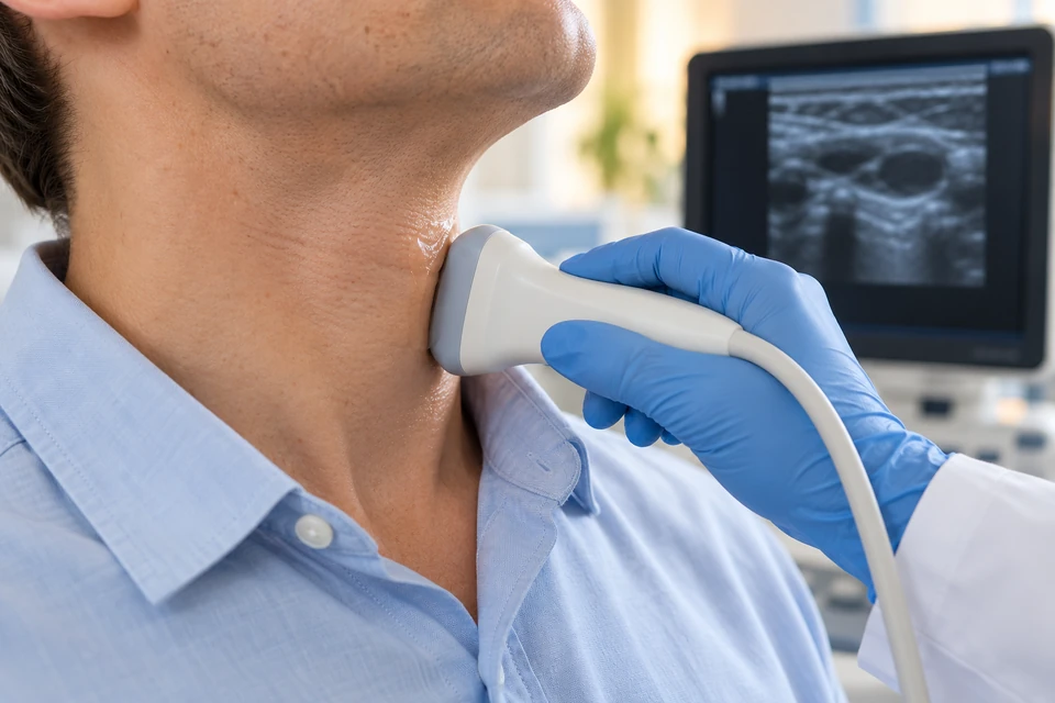
Assessment reviews duration, growth, pain, recent infection, medicines and symptoms such as fever, night sweats or unexplained weight loss. Blood tests and ultrasound are used when appropriate. Location affects access and nearby structures; the largest node is not automatically the best target. Previous cancer or biopsy results matter, and deep nodes may need image-guided sampling or another specialist approach.
Choosing needle or surgical biopsy
Fine-needle or core biopsy can provide sufficient information in selected situations with less intervention. Suspected lymphoma may require preserved architecture for diagnosis and subtyping, making excision useful when appropriate. Core biopsy can be considered if surgical risk is excessive. An inadequate sample does not exclude disease. The suspected diagnosis and pathology requirements should determine the method.
How excision is performed
A superficial node may be removed under local or general anaesthesia, depending on depth, location and circumstances. The surgeon accesses it through an incision, protects surrounding structures and controls bleeding. The specimen is sent for appropriate pathology, sometimes requiring advance laboratory coordination. The wound is closed and a drain is occasionally used. Visual appearance alone cannot establish the final diagnosis.
Location-specific risks
Risks include bleeding, bruising, infection, fluid or lymph collection, scarring and numbness. Nearby nerve and vessel risks differ between the neck, armpit and groin. Larger dissections have different lymphatic consequences from a single-node biopsy. Additional sampling or tests may still be needed. Anticoagulants and healing conditions are reviewed, and the expected scar should be discussed beforehand.
Preparation, aftercare and results
Share medicines, allergies and earlier biopsy reports, and do not stop anticoagulants independently. Fasting and home support depend on anaesthesia. Follow dressing, suture and drain instructions, with activity limits tailored to location. Pathology timing varies with required tests. Confirm who will explain the result and arrange subsequent care; a healed wound does not mean the diagnostic process is complete.
When prompt help is needed
Report rapidly increasing swelling, persistent bleeding, spreading redness, fever or offensive discharge. Breathing or swallowing difficulty after a neck procedure requires emergency care. New weakness, extensive numbness or severe pain needs assessment, as does increasing limb swelling after axillary or groin surgery. Fainting, chest pain or breathlessness are emergencies. Report further node enlargement or continuing systemic symptoms during follow-up.
This information is general. The cause of symptoms, appropriate investigations and treatment options are determined through individual assessment.