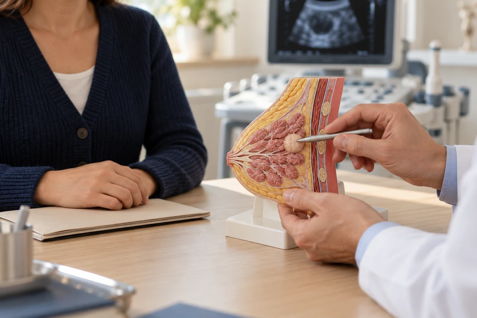
When is removal considered?
A palpable lump may be a cyst, fibroadenoma or another benign change, but touch alone cannot establish the diagnosis. Removal may be considered for growth, persistent discomfort, suspicious findings or unresolved uncertainty. The consultation should clarify what surgery is expected to achieve and whether another test could answer the question first.
Assessment before an operation
Assessment includes the lump's history, pregnancy or breastfeeding, previous procedures and family history. Ultrasound and mammography are selected according to the clinical situation. A core biopsy samples tissue when needed. Examination, imaging and pathology must agree; a benign biopsy result that does not explain a suspicious image may require further investigation.
Alternatives to excision
A confidently diagnosed, symptom-free benign lump may be observed. A painful simple cyst may be aspirated, while selected small lesions can sometimes be removed using an image-guided vacuum device. These approaches are not interchangeable. Diagnosis, location and symptoms determine suitability, and observation needs clear advice about changes that require reassessment.
How the operation is planned
Excision removes the target while preserving surrounding breast tissue where possible. An abnormality that cannot be felt may need image-guided localisation. Anaesthesia and incision placement depend on the planned operation. Suspected cancer requires a different margin strategy from a clearly benign lump, and the removed tissue is examined by a pathologist.
Risks and changes in appearance
Possible problems include bleeding, infection, fluid collection, discomfort and scarring. Removing a relatively large amount of tissue may change breast contour. Surgery near the nipple can affect sensation or milk ducts. Excision does not guarantee that unrelated breast pain will resolve, and an unexpected pathology result may lead to additional treatment.
Preparing for surgery
Bring earlier images, biopsy reports and a complete medication list. The team will advise about anticoagulants, diabetes treatment and supplements rather than asking you to stop medicines independently. Discuss allergies, smoking, previous anaesthetic problems and possible pregnancy. Fasting, transport and help at home depend on the type of procedure and anaesthesia.
Pathology and follow-up
Follow-up reviews both healing and the final diagnosis. The pathology must explain the imaging target, and further sampling or treatment may occasionally be necessary. Wound care and activity advice are individual. A new lump, nipple discharge or persistent change should be reported even when an earlier lump was benign.
When to seek prompt help
Contact the surgical team promptly for expanding swelling, persistent bleeding, spreading redness, fever or worsening pain. Breathlessness, chest pain or fainting require emergency assessment. Keep discharge contact details available and explain which procedure you had; the significance of symptoms depends on their progression as well as their intensity.
This information is general. The cause of symptoms, appropriate investigations and treatment options are determined through individual assessment.