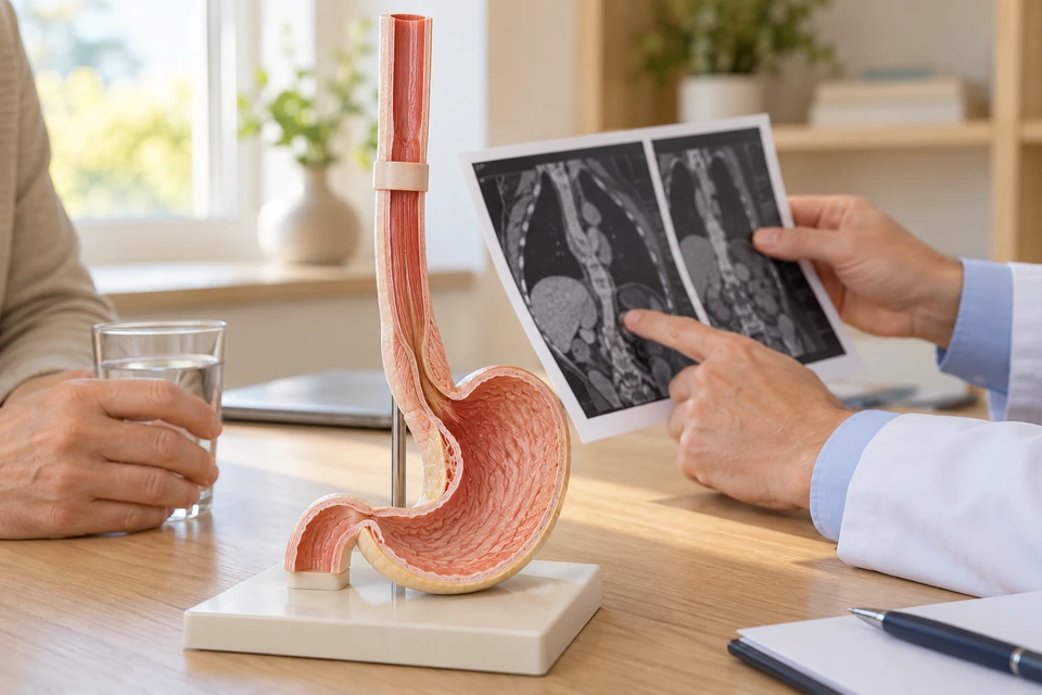
Where surgery fits into treatment
The oesophagus carries food to the stomach. Cancers at different levels and of different cell types do not share one treatment plan. Surgery may suit removable disease in a person fit for a major operation. Selected superficial tumours can be treated endoscopically, while other situations favour non-surgical treatment.
Diagnosis and assessment of spread
Endoscopic biopsy identifies the cancer. CT and, where appropriate, PET-CT or endoscopic ultrasound help assess its extent. Weight loss, swallowing and hydration are reviewed alongside heart and lung function. Symptom severity alone cannot determine stage or whether surgery is appropriate; imaging and pathology are essential.
Why treatment may involve several stages
Depending on stage and cell type, medicines or combined chemoradiotherapy may precede surgery. Definitive chemoradiotherapy can be an alternative in appropriate cases. Advanced disease may require systemic treatment and procedures that improve swallowing. The team should explain the purpose and sequence of each component, rather than presenting surgery in isolation.
How swallowing continuity is restored
The affected oesophagus and relevant lymph nodes are removed. Part of the stomach is commonly used to reconstruct the food passage, although alternatives may be needed. The join’s position and surgical access depend on tumour location and anatomy. Open and minimally invasive approaches are selected around safe cancer treatment.
Risks and support before surgery
Possible complications include leakage, chest infection, bleeding, clots, heart rhythm problems and later narrowing at the join. Nutrition and respiratory health deserve particular attention. Preparation may include dietetic support, alternative feeding, smoking cessation and exercise suited to ability. Medicines and the possible need for intensive care are also discussed.
Eating after the operation
Eating may involve temporary feeding support and gradual adjustment to the reconstructed passage. A dietitian helps plan smaller meals, protein and fluids. Early fullness, reflux or symptoms from rapid food transit may occur. Weight loss, food sticking or coughing during meals should be reported rather than accepted as inevitable.
Pathology and follow-up
Final pathology assesses treatment response, margins and lymph nodes, informing further care. Follow-up also reviews swallowing, nutrition, breathing and daily function. A narrowing may need endoscopic assessment and treatment. Supporting a workable eating routine and quality of life is part of follow-up alongside checking for cancer recurrence.
When to seek urgent help
Fever with worsening chest or abdominal pain, sudden breathlessness, fainting or rapid deterioration requires urgent assessment. Inability to swallow fluids, repeated vomiting or bleeding also needs prompt help. If a feeding tube is used, discharge instructions should explain whom to contact when it becomes displaced, blocked or otherwise problematic.
This information is general. The cause of symptoms, appropriate investigations and treatment options are determined through individual assessment.
Medical references
- NCI — Esophageal cancer treatment, professional PDQ
- NCI — Esophageal cancer treatment by stage
- NICE NG83 — Oesophago-gastric cancer recommendations
- University College London Hospitals — Oesophagectomy
- Cambridge University Hospitals — Eating after oesophagectomy
- American Cancer Society — Surgery for esophageal cancer