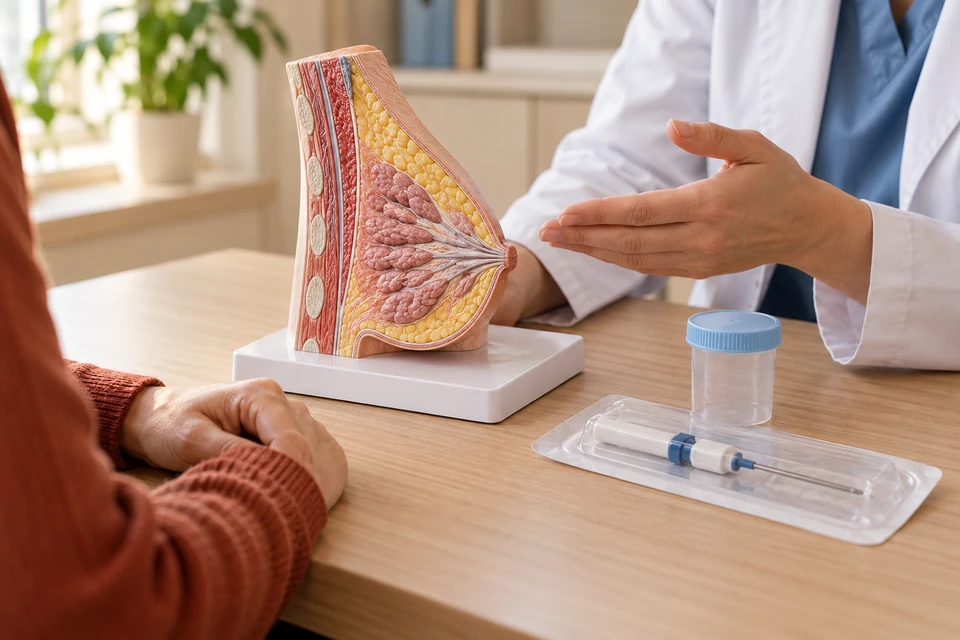
Why a biopsy is recommended
Examination or imaging may identify an area that needs tissue assessment. Biopsy helps distinguish benign changes, certain high-risk abnormalities and cancer. The target may be a lump, calcifications or an alteration that cannot be felt. Ask what finding is being sampled and how the result could change management.
Types of biopsy
Fine-needle aspiration obtains cells or fluid, while core biopsy preserves small pieces of tissue for structural assessment. Vacuum-assisted biopsy can collect more tissue. Surgical biopsy is reserved for selected circumstances. The method depends on the target and the diagnostic question; an inadequate sample may require another sampling approach.
Image guidance and targeting
Ultrasound, mammographic techniques or MRI may guide sampling, depending on how the abnormality is seen. A small marker may be left to identify the site later; its presence does not imply cancer. Keep the procedure report and marker information available for future imaging or treatment elsewhere.
Information needed before the procedure
Tell the team about medicines, supplements, bleeding problems, allergies, pregnancy and breastfeeding. Do not stop prescribed drugs independently. Preparation differs between a local-anaesthetic needle biopsy and a procedure involving sedation or surgery. Confirm fasting and transport arrangements, and tell the team if you experience pain or anxiety during sampling.
What happens during sampling?
The target is located, the skin prepared and local anaesthetic given when appropriate. Samples are taken through a small entry site and sent for pathology, followed by pressure and a dressing. Most needle biopsies are diagnostic rather than complete lump removal. Surgical biopsy has a different incision and anaesthetic plan.
Risks and aftercare
Bruising, tenderness and a small haematoma may occur. More substantial bleeding or infection is less common and depends partly on medicines and technique. Follow the procedure-specific dressing and activity instructions. Expanding tight swelling, persistent bleeding, worsening redness or fever requires prompt advice rather than being treated as ordinary bruising.
Why results must match the imaging
Pathology must be consistent with the sampled imaging abnormality and clinical findings. A benign result that does not explain a suspicious target may need repeat or surgical sampling. Some non-cancerous findings also need further assessment. The results discussion should address adequacy, diagnostic agreement and the next recommended step.
Clarifying the next step
Before leaving, confirm how the results will be discussed. Additional laboratory tests can affect timing. Depending on the findings, the next step may be routine screening, surveillance, further sampling or treatment planning. Keep copies of reports and images. Attending the results discussion completes the diagnostic process.
This information is general. The cause of symptoms, appropriate investigations and treatment options are determined through individual assessment.