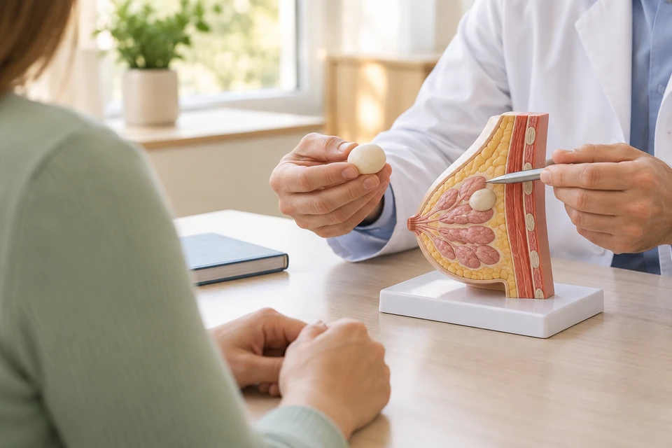
Fibroadenoma and cancer
A fibroadenoma is a benign growth containing glandular and supporting tissue. It may feel smooth and mobile, but those features alone are not diagnostic. New or changing lumps still require assessment. Several fibroadenomas can coexist, and an established diagnosis does not mean that every later breast change has the same cause.
Establishing a reliable diagnosis
Examination and age-appropriate imaging are considered together, often beginning with ultrasound. Needle biopsy is used when indicated. A fibroadenoma result is most reassuring when it matches the imaging. Rapid growth or discordant findings may require further sampling to distinguish another fibroepithelial lesion, including a phyllodes tumour.
When can observation be enough?
A symptom-free fibroadenoma with concordant imaging and pathology may not need removal or repeated short-interval scans. Return to age- and risk-appropriate screening can be considered. The plan depends on diagnostic confidence and clinical circumstances. Observation should include clear instructions about growth or other changes that warrant another review.
Deciding whether to remove it
Removal may be discussed for substantial growth, distortion, persistent symptoms, uncertainty or informed preference. One size threshold does not decide every case. Age, pregnancy and breastfeeding affect interpretation. Surgery may not resolve pain from another cause, so the expected benefit should be weighed against scarring, sensation changes and tissue loss.
Surgical and other removal methods
Surgical excision removes the lump while preserving surrounding tissue where possible, followed by pathology examination. Incision placement depends on anatomy. Image-guided vacuum-assisted removal may suit selected small lesions but is not appropriate for every fibroadenoma. Compare anaesthesia, completeness of removal and the information each approach provides.
Risks and preparation
Risks include bruising, bleeding, infection, fluid collection, scars and contour changes. Procedures near the nipple may affect sensation or milk ducts. Bring imaging and biopsy records and discuss medicines, allergies, smoking and pregnancy. Medication changes and transport arrangements should follow the agreed anaesthetic and surgical plan.
Pathology and subsequent follow-up
Pathology confirms the diagnosis and may occasionally identify a different fibroepithelial lesion requiring further assessment. Recovery advice depends on the procedure. Removing one fibroadenoma does not prevent another from developing elsewhere. A lump at the operation site can also represent scar or fluid, so new findings should be assessed rather than assumed.
When should you seek earlier review?
Seek earlier review for rapid growth, skin dimpling, bloodstained nipple discharge or a new axillary swelling. After removal, increasing pain, fever, spreading redness or expanding swelling needs prompt advice. At follow-up, ask whether pathology confirmed the expected diagnosis and what screening or reassessment is appropriate for the future.
This information is general. The cause of symptoms, appropriate investigations and treatment options are determined through individual assessment.