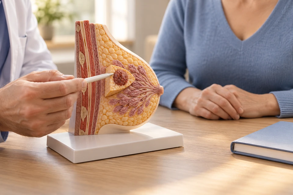
What does lumpectomy mean?
Lumpectomy describes removal of a defined breast lesion. For cancer, it aims to remove the tumour with suitable margins while preserving most of the breast. Similar terminology is sometimes used for benign lump removal. Clarifying whether the purpose is diagnosis or cancer treatment helps explain the operation's extent and the follow-up required.
How is the target identified?
A palpable lump may not represent the full imaging abnormality. Small lesions, calcifications and tumours reduced by preoperative treatment may require localisation using a wire or another marker. Localisation identifies the target; it does not replace assessment of disease extent. The imaging and biopsy must refer to the same lesion.
Suitability and alternatives
Suitability depends on disease distribution, tumour-to-breast size and the feasibility of radiotherapy. Another conserving technique or mastectomy may be discussed if adequate margins or an acceptable contour are unlikely. Preoperative medicines may alter the options. Compare the complete treatment pathway rather than judging procedures only by incision size.
The main steps
The target and planned surrounding tissue are removed under anaesthesia. The specimen is oriented for pathology and may be imaged when needed. Lymph-node assessment, if indicated, follows a separate plan. Closing the tissue depends on the defect. The final microscopic assessment remains important even when removal appears complete during surgery.
Could another operation be needed?
Pathology examines the tumour and its relationship to the margins. Further excision, or sometimes mastectomy, may be advised when clearance is inadequate. Requirements differ with the type of disease. A second operation does not automatically indicate an error: microscopic spread may only become apparent after the entire specimen has been examined.
Risks and breast appearance
Bleeding, infection, fluid collection, scars and altered sensation are possible. Volume loss may create a dent or asymmetry, and radiotherapy can produce additional changes. Oncoplastic options may be relevant before surgery. Axillary procedures require separate advice about movement and lymphoedema; removing a lesion does not guarantee relief of unrelated breast pain.
Preparation and daily life
Localisation appointments may need coordination with the operation. Follow individual instructions about fasting, medicines and anaesthesia. At home, use the advised wound care and activity plan. Driving, work and exercise depend on comfort, movement and any additional procedures. The follow-up appointment should include pathology discussion, not just a wound check.
Next treatment and prompt review
Radiotherapy is commonly considered after cancer lumpectomy, while systemic treatment depends on tumour features. Follow-up imaging remains important. Seek prompt help for expanding swelling, persistent bleeding, fever or spreading redness. Report a new lump or bloodstained nipple discharge. The results appointment should clarify margin adequacy and the next treatment step.
This information is general. The cause of symptoms, appropriate investigations and treatment options are determined through individual assessment.