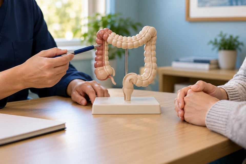
Right and left hemicolectomy
Hemicolectomy removes a defined side of the colon; it does not mean exactly half is removed in every case. Right hemicolectomy involves the caecum and ascending colon region, while left hemicolectomy removes an affected left-sided segment. Extended resections may be needed. Indications include cancer, selected polyps unsuitable for endoscopic removal, impaired blood supply and other localised disease.
Investigations that define the operation
Colonoscopy and biopsy help locate and characterise the lesion, while imaging assesses extent, especially for cancer. Some small lesions may need endoscopic marking. Cancer-related blood vessels and lymphatic drainage influence resection boundaries. Previous bowel surgery, kidney function, nutrition and anaemia are reviewed. Recording usual bowel habits helps later comparison. Uncertainty about the lesion's position should be addressed before planned surgery.
Alternatives and shared decisions
Advanced endoscopic treatment may suit some benign polyps, and medicines may help selected inflammatory conditions. These are not appropriate substitutes for every tumour or complication. A limited resection versus hemicolectomy is chosen around adequate disease treatment and reliable blood supply. Cancer surgery may need regional lymph nodes beyond the visible lump. Decisions should address diagnostic certainty, fitness and the disease-specific consequences of waiting.
How the bowel is reconnected
After right hemicolectomy, the end of the small bowel may be joined to the remaining colon. Left-sided surgery joins the remaining colonic segments or an appropriate lower segment. A stoma may be needed if a safe connection is not possible. Open or minimally invasive surgery is selected individually. The final pathology examines the removed specimen and helps determine whether further treatment is required.
Risks and bowel changes
Risks include leakage, infection, bleeding, delayed bowel activity, adhesional obstruction and blood clots. Nearby structures and the bowel segments removed create additional individual considerations. Stool consistency or frequency may change. Persistent diarrhoea after right-sided surgery can have treatable causes and should not automatically be labelled adaptation. Assessment may consider diet, medicines, infection and absorption before deciding on management.
Preparation and reviewing results
Preparation addresses anaemia, nutrition, smoking and suitable exercise, with personalised medication and bowel preparation instructions. Recovery combines pain relief, movement and a return to eating. Monitor the wound, hydration and bowel pattern at home. For cancer, final pathological staging informs additional treatment and surveillance. Follow-up should also cover bowel symptoms, work, exercise and practical concerns, rather than only the operation's technical result.
When prompt assessment is needed
Increasing pain, fever, distension, persistent vomiting or worsening wellbeing should be reported promptly. Failure to pass stool or gas is concerning when accompanied by pain. Wound pus, opening or spreading redness need assessment. Watery stools with reduced urine or dizziness suggest fluid loss. Chest pain, breathlessness, fainting or heavy bleeding may require emergency care, even after an initially uncomplicated discharge.
This information is general. The cause of symptoms, appropriate investigations and treatment options are determined through individual assessment.